The human nervous system is the most complex structure known to science. It governs everything from our physical movements and sensory perceptions to our thoughts, memories, and core personality traits. When disease, trauma, or structural defects threaten this delicate system, patients rely on a highly specialized medical expert: the neurosurgeon.
A neurosurgeon is a medical doctor who diagnoses, evaluates, and treats disorders of the central and peripheral nervous systems. This includes the brain, spinal cord, skull, vertebral column, and the intricate network of nerves that extends to the tips of the fingers and toes. While many people equate neurosurgery exclusively with brain surgery, a significant portion of a neurosurgeon’s daily practice actually focuses on spinal disorders and nerve reconstructions.
Understanding the role of these physicians requires looking into their intensive educational path, the diverse clinical conditions they treat, the cutting-edge technologies they utilize, and the realities of managing patient care in a high-stakes environment.
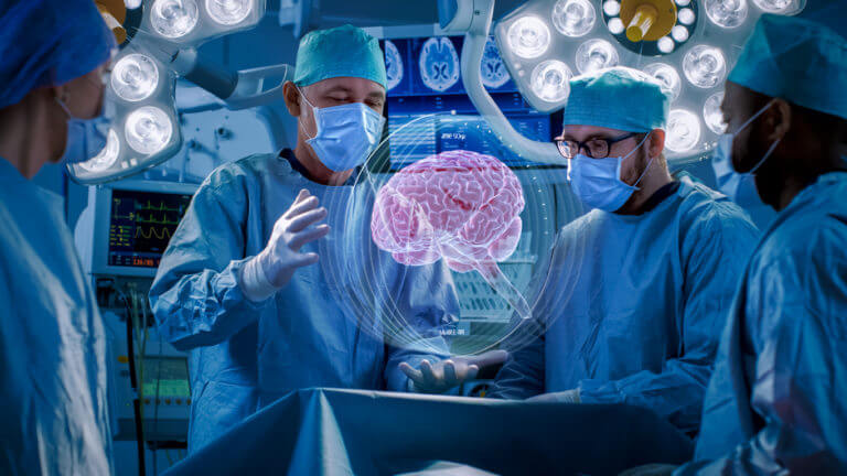
The image above highlights the highly technical environment where modern neurosurgeons operate. Notice the presence of digital displays, high-powered microscopes, and robotic guidance systems. These tools work together to allow physicians to visualize microscopic pathways and navigate the delicate anatomy of the brain and spine with millimeter precision.
The Academic and Clinical Training Journey
Becoming a neurosurgeon demands one of the longest and most rigorous training pathways in all of medicine. The journey requires an immense commitment of time, intellectual energy, and physical stamina.
-
Undergraduate Degree: Aspiring physicians complete a four-year bachelor’s degree, typically focusing on pre-medical sciences such as biology, chemistry, or neuroscience, while maintaining an exceptional academic record to secure admission to medical school.
-
Medical School: Students spend four years completing a Doctor of Medicine or Doctor of Osteopathic Medicine program. During this period, they acquire foundational medical knowledge and participate in clinical rotations across various specialties to develop a comprehensive understanding of patient care.
-
Neurosurgery Residency: Upon graduation, new doctors must match into a dedicated neurosurgery residency program. In the United States, this residency lasts seven years. This intensive period constitutes the core of their practical training. Residents work under direct supervision, gradually advancing from basic post-operative care to executing complex surgical procedures.
-
Fellowship Specialization: Many neurosurgeons choose to spend an additional one to two years completing a specialized fellowship. This advanced training focuses on subspecialties such as pediatric neurosurgery, neurovascular intervention, neuro-oncology, or complex spine surgery.
By the time a neurosurgeon begins practicing independently, they have typically completed a minimum of fourteen to fifteen years of higher education and clinical training. This lengthy preparation ensures they possess the refined technical skills and rapid decision-making abilities necessary to navigate life-or-death scenarios in the operating room.
Core Clinical Focus Areas and Procedures
The scope of neurosurgery extends far beyond a single organ. Neurosurgeons manage conditions that impact the structural integrity and functionality of the entire nervous system. Their clinical responsibilities are generally divided into several key areas.
Cerebrovascular Surgery
This subspecialty focuses on the blood vessels supplying the brain and spinal cord. Neurosurgeons treat conditions like brain aneurysms, which are weakened spots in arterial walls that risk rupturing and causing life-threatening hemorrhagic strokes. They also address arteriovenous malformations, which are abnormal tangles of blood vessels that disrupt normal circulation. Procedures include open craniotomies to place tiny metal clips across an aneurysm neck, as well as endovascular techniques where long, thin catheters are guided through the blood vessels from the groin or wrist to seal off abnormalities from the inside.
Neuro-Oncology
Neuro-oncologists specialize in the surgical management of primary and metastatic tumors affecting the brain, spinal cord, and surrounding membranes. Primary tumors, such as glioblastomas or meningiomas, originate directly within the skull, whereas metastatic tumors spread from cancers located elsewhere in the body. The primary goal of the neurosurgeon is to safely maximize the extent of tumor removal while meticulously preserving the surrounding, functional brain tissues responsible for movement, speech, and cognition.
Spine Neurosurgery
Spinal procedures make up more than half of the total surgeries performed by many neurosurgeons. They address degenerative disc diseases, herniated discs, spinal stenosis, spinal deformities like scoliosis, and traumatic vertebral fractures. When a disc or bone fragment compresses the spinal cord or exiting nerve roots, it can cause severe pain, numbness, or progressive paralysis. Neurosurgeons perform decompressions, laminectomies, and complex spinal fusions using hardware like rods, screws, and artificial cages to restore structural stability and alleviate nerve pressure.
Functional and Stereotactic Neurosurgery
This field addresses conditions where the structure of the brain appears normal, but its electrical circuitry functions abnormally. It includes the surgical management of epilepsy, severe chronic pain, and movement disorders such as Parkinson’s disease and essential tremor. A prominent procedure in this domain is Deep Brain Stimulation, where the surgeon implants thin electrodes into specific, deep brain structures. These electrodes connect to an internalized pulse generator that delivers targeted electrical currents to modulate abnormal signaling pathways and restore a patient’s motor control.
Pediatric Neurosurgery
Children experience a distinct set of neurological disorders compared to adults. Pediatric neurosurgeons treat congenital anomalies such as spina bifida, craniosynostosis (premature fusing of the skull bones), and hydrocephalus (an abnormal accumulation of cerebrospinal fluid within the brain cavities). Managing pediatric patients requires specialized techniques tailored to growing skeletal structures and delicate, developing nervous tissues.
Advanced Technological Innovations
The field of neurosurgery evolves in close alignment with technical innovation. Over the past several decades, advancements in engineering, computer science, and imaging have radically altered surgical protocols, drastically reducing patient risk and improving recovery trajectories.
Image-Guided Neuronavigation
Modern neurosurgeons utilize advanced computer systems that function much like a global positioning system for the human body. Before an operation, high-resolution magnetic resonance imaging and computed tomography scans are uploaded into a navigation workstation. In the operating theater, infrared cameras track the surgeon’s specialized instruments in real time, projecting their exact position onto a three-dimensional model of the patient’s brain. This capability allows the physician to locate deep-seated pathologies through the smallest, safest corridor possible, minimizing unnecessary disruption to healthy brain architecture.
Minimally Invasive Approaches
Traditional skull and spinal surgeries historically required extensive incisions and substantial muscle retraction. Today, minimally invasive techniques utilize narrow tubes, specialized endoscopes, and high-definition cameras to access operative sites through keyhole openings. For example, many pituitary gland tumors are now accessed entirely through the nasal passages using an endoscope, avoiding any visible external scars or direct incisions into the skull. In spine surgery, minimally invasive techniques preserve muscle attachments, which directly translates to less postoperative pain, reduced blood loss, and a accelerated return to daily activities.
Intraoperative Mapping and Monitoring
To safely remove tumors or epileptic tissue located near critical functional hubs, such as the motor cortex or language centers, neurosurgeons frequently employ intraoperative neuromonitoring. This technique involves placing electrodes on the patient’s skin or directly on the surface of the nervous tissue to monitor electrical pathways during the procedure. In specific instances, surgeons perform awake brain surgery. During these operations, the patient is gently awakened midway through the procedure to participate in language or motor tests while the surgeon maps the borders of a tumor, ensuring vital functional pathways remain completely unharmed.
The Realities of Daily Practice
The life of a neurosurgeon is characterized by intense intellectual engagement balanced against profound physical and emotional demands. A typical week involves a mix of diagnostic consultations in an outpatient clinic, multi-disciplinary review panels with oncologists and radiologists, hours spent standing under the heavy magnification lamps of the operating room, and managing critical care recovery in the neuro-intensive care unit.
Because neurological emergencies can occur at any hour, neurosurgeons frequently maintain demanding on-call schedules. They are routinely called upon to perform emergency procedures for acute traumatic brain injuries, ruptured aneurysms, or sudden spinal cord compressions resulting from accidents. Delivering care in these environments requires immense emotional resilience, a steady hand under pressure, and the clear communication skills needed to help patients and their families navigate sudden, life-altering medical crises.
Frequently Asked Questions
What is the difference between a neurologist and a neurosurgeon?
A neurologist is a medical specialist who diagnoses, manages, and treats neurological disorders primarily through non-surgical means, such as medication, physical therapy, and lifestyle adjustments. In contrast, a neurosurgeon is trained to provide both conservative management and surgical interventions when a condition requires manual repair, tissue removal, or structural stabilization of the nervous system.
How do neurosurgeons minimize the risk of permanent brain damage during surgery?
Neurosurgeons rely on a multi-layered framework of safety technologies. This includes real-time image guidance systems, continuous intraoperative electrophysiological monitoring of nerve pathways, and fluorescence-guided surgery to differentiate tumor margins from normal tissue. When necessary, they perform awake mapping to actively test speech and motor skills during the operation.
Why do neurosurgeons treat spinal conditions instead of orthopedic surgeons?
Both neurosurgeons and orthopedic surgeons perform complex spine surgeries, and both complete extensive training in spinal anatomy. However, neurosurgeons are uniquely trained throughout their entire residency to open and operate inside the dura mater, which is the protective membrane enclosing the spinal cord and nerve roots, making them highly specialized in managing the neural elements themselves.
What is a craniotomy and is the patient always awake during it?
A craniotomy is a standard surgical procedure where a section of the skull bone is carefully removed to give the neurosurgeon access to the brain beneath. The bone segment is securely replaced and fastened at the end of the operation. The vast majority of craniotomies are performed under general anesthesia while the patient is completely asleep; awake craniotomies are utilized exclusively when a tumor or lesion sits directly adjacent to essential speech or movement centers.
What causes hydrocephalus and how do neurosurgeons treat it?
Hydrocephalus occurs when there is an imbalance between the production and absorption of cerebrospinal fluid, or when the normal flow of this fluid through the brain’s internal chambers is blocked, causing dangerous pressure build-up. Neurosurgeons typically treat this by implanting a shunt system, which is a flexible tube that diverts excess fluid from the brain down into another part of the body, such as the abdomen, where it can be safely reabsorbed.
What does the recovery timeline look like after major spinal surgery?
The recovery period varies substantially depending on the specific procedure performed and the patient’s baseline health. Following minor or minimally invasive spinal decompressions, patients are often able to return home the same day or after a brief overnight stay, resuming light office work within a few weeks. Major spinal fusions, however, typically require several days of inpatient monitoring followed by several months of structured physical therapy to allow the bone graft to fully solidify and stabilize.